Novembers Case of the Month-2023
Lymphoma with Focal Renal Lesion
Signalment
Age: 9 years 6 months
Gender: Female Spayed
Species: Feline
Breed: Domestic Shorthair (DSH)
Weight: 6 lbs
History
The patient presented with weight loss. Abdominal palpation revealed irregularities in kidney shape and a suspected abdominal mass. An abdominal ultrasound was requested to further evaluate these findings and investigate potential causes of the weight loss.
Ultrasound Findings and Images
Kidneys: The kidneys are of normal size (Left: 3.9 cm, Right: 4.0 cm) with a normal to mildly elongated shape. The cortical and medullary echotexture is heterogeneous and hypoechoic. Trace to mild hypoechoic subcapsular fluid is present. A hypoechoic, mildly vascular, rounded, and well-defined nodule (1.1 x 1.0 cm) is protruding from the cranial pole of the right kidney, with an intact capsule. No pyelectasia was observed.
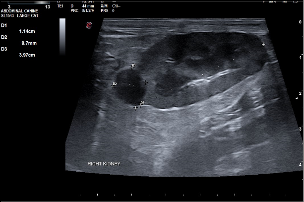
Image 1: Right kidney showing a cranial pole capsule-altering hypoechoic nodule with trace subcapsular fluid.
Bladder
The bladder is moderately distended with mildly echogenic urine containing amorphous suspended debris. Its contour and thickness are relatively normal. No overt obstruction, uroliths, or neoplasia noted.
Small Intestine
Many loops of the intestine are prominent and mildly to severely thickened:
- Duodenum: 3.1–3.7 mm
- Jejunum: 2.7–4.5 mm
- Ileum: 6.4 mm
Normal intestinal wall thickness is approximately 2.2–2.5 mm, with >2.8 mm considered abnormal (Norsworthy/Estep et. al., JAVMA, Vol 243, No. 10, November 15, 2013). Abnormal layering with prominent disproportionate thickening of the muscularis layer is observed.
Lymph Nodes
Multiple mesenteric lymph nodes are severely enlarged with a rounded shape and homogenous hypoechoic echogenicity (~3.5 x 2.3 cm). The ICCJ lymph nodes are mildly hypoechoic and plump (~0.7 cm).
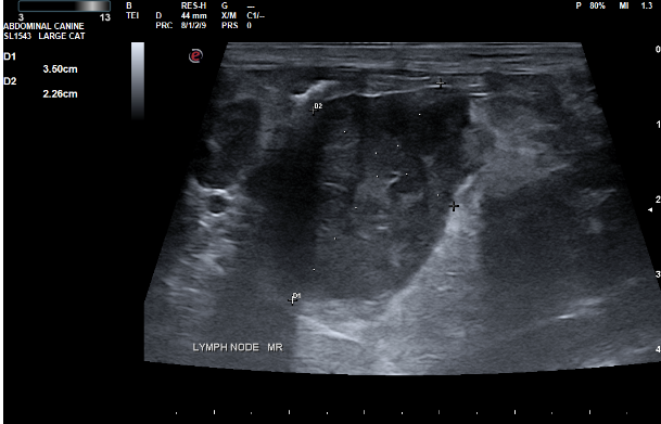
Image 2
Description: Enlarged hypoechoic heterogeneous mesenteric lymph node with hyperechoic mesentery.
Serosal Surfaces
- Findings: Trace amount of free fluid in the abdomen. Mesentery throughout the abdomen is diffusely hyperechoic.
Abdominal Ultrasound Interpretation
Lymph Nodes
- Severity: Severe
- Differential Diagnoses (DDx):
- Infiltrative neoplasia (lymphoma, mast cell tumor, other)
- Inflammatory bowel disease (IBD)
- Infection or reaction
- Metastatic neoplasia
Kidneys
- Severity: Mild to moderate
- Differential Diagnoses (DDx):
- Chronic nonspecific changes (chronic glomerulonephritis, amyloidosis, chronic interstitial nephritis)
- Acute renal failure or nephritis (infectious, glomerulonephritis, toxic, etc.)
- Acute-on-chronic renal failure
- Lymphosarcoma
- Pyelonephritis
Right Kidney Nodule
- Severity: Moderate
- Differential Diagnoses (DDx):
- Renal lymphosarcoma (diffuse or focal mass)
- Primary renal carcinoma or transitional cell carcinoma (TCC)
- Renal malignant histiocytosis or mastocytosis
- Malignant osteosarcoma or hemangiosarcoma (metastatic)
- Fungal infection (Cryptococcus, Aspergillosis)
Intestines
- Severity: Mild to severe
- Differential Diagnoses (DDx):
- Inflammatory bowel disease (IBD) or food intolerance
- Infiltrative neoplasia (small-cell lymphosarcoma, mast cell tumor)
- Parasitism (e.g., cestodes)
- Dry feline infectious peritonitis (FIP)
- Fungal infection (Histoplasmosis)
Ascites
- Severity: Mild
- Differential Diagnoses (DDx):
- Transudate
- Hemorrhagic fluid
- Exudate
Mesentery
- Severity: Mild to moderate
- Differential Diagnoses (DDx):
- Peritonitis (inflammation)
- Paraneoplastic reaction
- Infectious process
- Fibrosis
- Other
Echogenic Urine
- Differential Diagnoses (DDx):
- Cellular debris
- Lipids
- Proteins
- Amorphous debris
Further Testing
Ultrasound-guided freehand biopsies of the mesenteric lymph node and the kidney nodule were collected for cytology.
Cytologic Diagnosis and Findings
Kidney Nodule
- High-grade granular lymphoma
Mesenteric Lymph Node
- High-grade granular lymphoma
Diagnosis: High-grade lymphoma is definitive (100% confidence) for both locations. The granular morphology suggests lymphocytes of cytotoxic T-cell or possibly NK-cell origin.
Microscopic Description
The slides from both locations are similar, moderately to highly cellular, and consist of:
- Few red blood cells
- Nucleated cells predominated by large immature lymphocytes, with few small lymphocytes and occasional neutrophils
- Large lymphocytes with:
- Basophilic cytoplasm occasionally containing small pink granules
- A perinuclear clear area
- Large round nucleus with finely-stippled chromatin and 1–3 prominent nucleoli
- Occasional mitotic figures noted
- Background consists of lymphoglandular bodies and occasional free nuclei
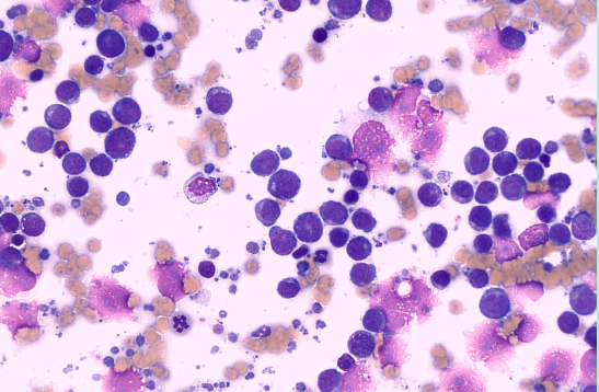
Image 3
Kidney: Numerous large immature lymphocytes
Discussion
Lymphoma is a commonly found neoplasia in feline patients. These patients may present with vague clinical signs such as weight loss and malaise. The incidence of renal involvement in feline lymphoma patients is estimated to be 3.6% to 30%.
Multicentric lymphoma is suspected in this patient due to a focal renal lesion (rather than bilateral renal enlargement) and lymph node involvement with intestinal changes suggestive of lymphoma.
Outcome
The referring veterinarian and patient owner are performing additional baseline diagnostics and discussing options for treatment.
References
Debruyn K, Haers H, Combes A, et al. Ultrasonography of the feline kidney: Technique, anatomy and changes associated with disease. Journal of Feline Medicine and Surgery. 2012; 14 (11): 794-803. doi: 10.1177/1098612X12464461
Valdés-Martínez A, Cianciolo R, Mai W. Association between renal hypoechoic subcapsular thickening and lymphosarcoma in cats. Vet Radiol Ultrasound. 2007 Jul-Aug;48(4):357-60. doi: 10.1111/j.1740-8261.2007.00256.x. PMID: 17691636.
The Curbside guide- Diagnosis & Treatment of Common Sonographically Detected Disease: Canine & Feline; Lindquist/ Frank/ Modler Lobetti, 2015, Sonopath LLC., pp. 181
Williams AG, Hohenhaus AE, Lamb KE. Incidence and treatment of feline renal lymphoma: 27 cases. J Feline Med Surg, 2021 Oct;23 (10):936-944. doi: 10.1177/1098612X20984363. Epub 2021 Jan 19. PMID: 33464143
Jeon J, Song D, Ro W, Kim H, Lee G, Cho J, Jeong W, Kim S, Sur J, Park H. Renal Lymphoma with Mesenteric Lymphomatosis in a Cat. J Vet Clin 2020;37:208-212. https://doi.org/10.17555/jvc.2020.08.37.4.208
Thank you Hope Animal Hospital and Eastern Vet Path for collaborating with us on this case!