Thoracic Disease in a Dog
Patient Information:
Age: 2 years old
Species: Canine
Breed: Standard Poodle
Gender: Spayed Female
History: The patient presented with lethargy, fever, and mild increased respiratory effort. She has been receiving prednisone and cyclosporine for immune-mediated thrombocytopenia diagnosed 6 months prior. Bloodwork showed mild thrombocytopenia, leukocytosis, normal chemistry, and negative 4DX. The patient exhibited a poor response to initial therapy with enrofloxacin.
Thoracic radiographs: Possible cranial thoracic lymphadenopathy. Otherwise, normal thorax.
Thoracic ultrasound: Multiple well-defined circular hypoechoic and slightly heterogeneous hyperechoic nodules were observed in the cranial mediastinal space. The lesions varied in size, the largest measuring 2.0x3.4cm in diameter. The septum and connective tissue appeared severely hyperechoic and a moderate amount of echogenic fluid was present in the cranial mediastinal space.
Image # 1: Ultrasound video depicting multiple fungal lesions of varying size and echogenicity in the cranial mediastinal space.
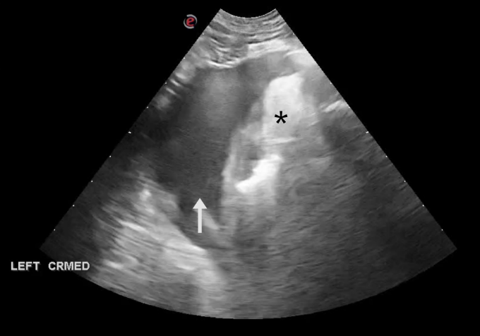
Images #2: Ultrasound image showing a moderate amount of cellular pleural effusion (arrow) and inflammation of the connective tissue (asterisk) of the cranial mediastinum.
Cytology: Ultrasound-guided thoracocentesis and fine-needle biopsies of the nodular lesions were performed under sedation. Gross assessment of the samples revealed black-pigmented deposits. Cytology results from both sites revealed marked pyogranulomatous inflammation with fungal hyphae consistent with Phaeohyphomycosis. Additional diagnostics recommended included PCR testing and fungal culture and sensitivity.
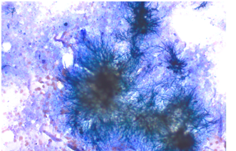
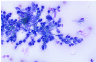
Left image (nodule) – Right image (fluid): Present across the slides were mixed inflammatory cells and variably intact aggregates of green-pigmented fungal hyphae. These hyphae displayed parallel walls, septations, and acute right-angle branching with rounded to tapered terminations consistent with Phaeohyphomycosis.
Comments: Phaeohyphomycosis is a rare fungal infection caused by dematiaceous (darkly pigmented) fungi containing melanin in their cell walls. These fungi are commonly found in soil, plants, and decaying organic matter. Phaeohyphomycosis has been reported in cats, dogs, cows, and horses, primarily affecting the skin and subcutaneous tissues, but the infection can become systemic involving the lungs and central nervous system. Patients treated with multiple immunosuppressive agents, particularly cyclosporine, appear at higher risk. Diagnosis is confirmed via cytology, histopathologic, fungal culture, and PCR assay. Treatment involves discontinuing immunosuppressive therapy. Surgical excision may be required for localized lesions. Non-resectable disease is treated with itraconazole, voriconazole, or posaconazole; however, the prognosis remains poor due to limited antifungal efficacy.
- Todoroff RJ, Lloyd DH. Approaches to opportunistic fungal infections in small animals. Today’s Veterinary Practice [Internet]. Merck Veterinary Manual. Fungal infections in dogs. Merck Veterinary Manual [Internet].
- Perfect JR, Schell WA. The new fungal opportunists are coming. Clinical Infectious Diseases. 2002;38(3):e15-e20. doi:10.1086/366054.
- Seyedmousavi S, Guillot J, de Hoog GS. Phaeohyphomycosis, emerging fungal diseases in animals. Frontiers in Veterinary Science [Internet]. 2019;6:249. doi:10.3389/fvets.2019.00249.
Outcome: due to disease progression, poor prognosis, and declining quality of life, the owner elected for humane euthanasia.
Special thanks to Dr. Palmar and the staff at Palmar Animal Hospital for their help with this case.