Unusual presentation of Renal Neoplasia in a Feline
Patient Information
Age: 8 years
Gender: Spayed Female
Breed: DSH
Species: Feline
History
Patient presented for weight loss, increased respiratory effort, and constipation.
Abdominal Ultrasonographic Findings
The left kidney is moderately enlarged (4.6cm length) with an abnormal shape. The kidney has a hyperechoic cortex with normal corticomedullary dimension distinction and is surrounded by a wide band of hypoechoic tissue. Vascularity is absent on Doppler exam. Moderate diverticular mineralization is present. No significant pyelectasia noted.
There is a small amount of retroperitoneal fluid present, and the retroperitoneal tissue is moderately hyperechoic around the left kidney.
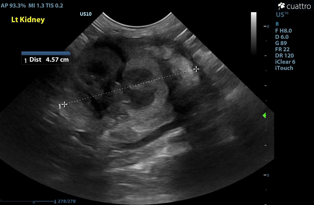
Image of left kidney. An irregularly shaped kidney surrounded by a large band of hypoechoic tissue.
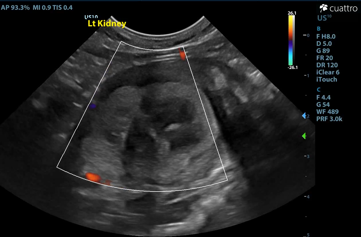
Doppler exam showing absent blood flow to organ.
Ultrasound Interpretation
Left kidney: The findings are severe. Differential diagnoses include complete renal infarct, torsion, strangulation, necrotic neoplasia, or severe nephritis.
Retroperitoneal space: The findings are moderate. Differential diagnoses include peritonitis (inflammation vs. infectious), fibrosis, or metastatic neoplasia.
Diagnostic Recommendations
Given the severity of changes to the left kidney and the obvious deleterious effect on the patient’s condition, exploratory laparotomy was recommended with excisional biopsies.
Histopathology Results
Diagnosis: Renal Cell Carcinoma, highly pleomorphic with invasion into peritoneal adipose tissue and severe and extensive necrosis.
Patient Outcome
The abnormal kidney was firmly adhered to the body wall. The decision was made that safe removal would be difficult and, therefore, humane euthanasia was elected at the time of surgery. Biopsy and histopathology were performed for professional interest.
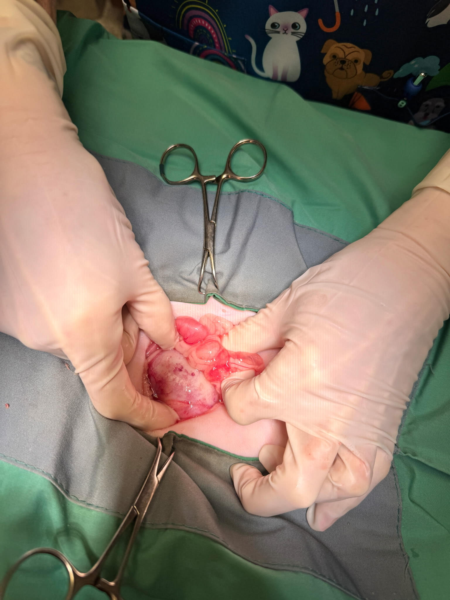
Image of kidney prior to removal Cross section of left kidney.
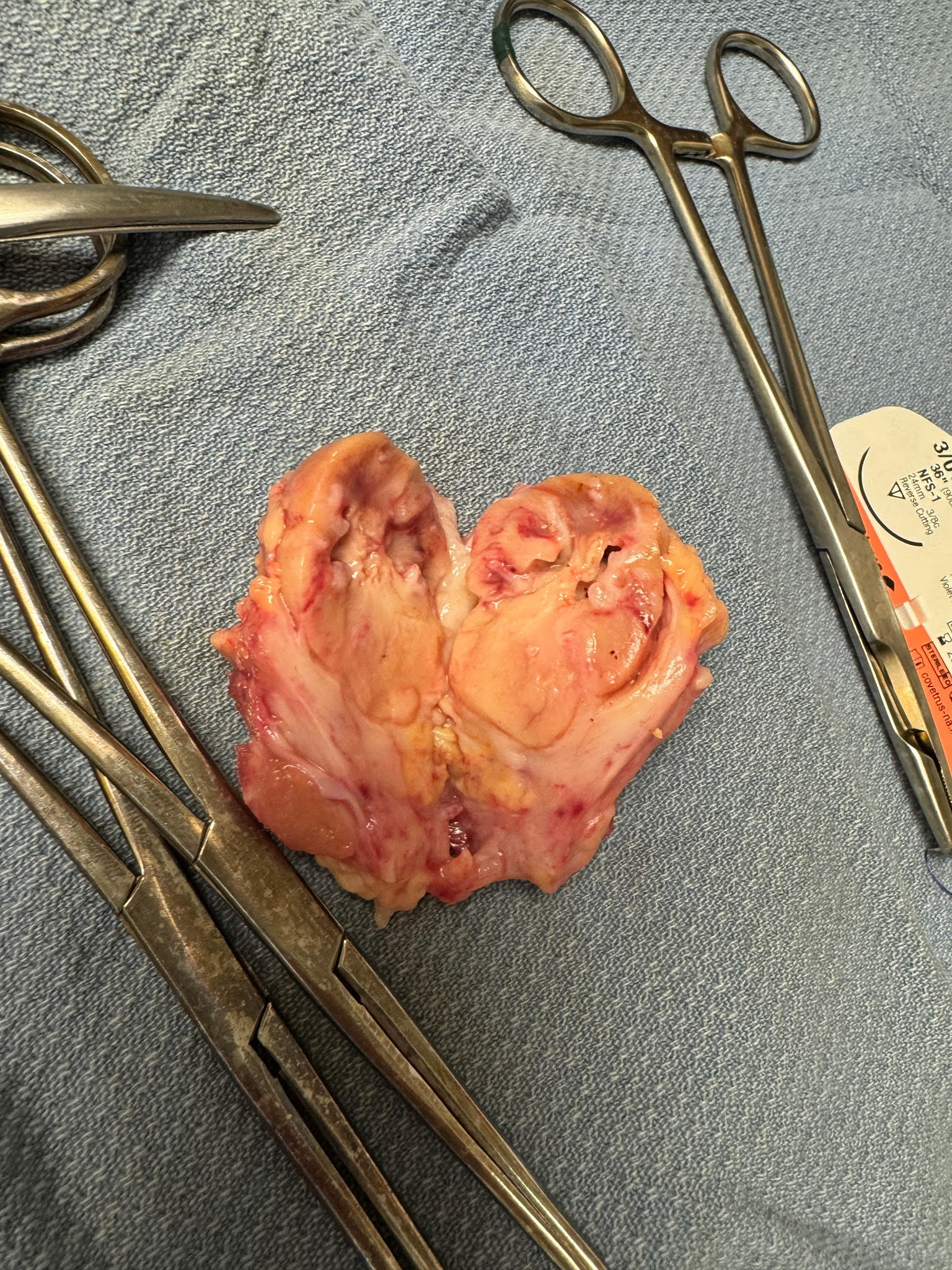
The kidney is surrounded by a large amount of fibrous tissue.
Discussion
Lymphoma is the most common neoplasia of the kidneys in cats. Primary renal tumors are far less common in this species, and limited peer-reviewed literature is available. 77% of primary renal neoplasia is epithelial in origin in cats, with a 50-64% metastasis rate. Common metastasis sites are the lungs, liver, regional lymph nodes, and adrenal glands. Surgery is the treatment of choice since most tumors are unilateral.
In one recent study, the mean survival time was 1217 days following nephrectomy (in patients that survived to discharge). In this particular case, the carcinoma had diffusely affected the renal tissue rather than forming a focal mass and created significant necrosis, which had compromised the blood supply, creating a clinical situation similar to torsion or complete renal infarct. Significant adhesion to surrounding tissue had occurred.
References
- Kenny et al. 2023. Clinical outcomes of cats with renal carcinoma undergoing nephrectomy: A retrospective study. Veterinary and Comparative Oncology. 2023; 21(4): 565-748.
- Meuten. 2017. Tumors of Domestic Animals. 5th ed. pp 638-644.
- Matsumoto et al. Histopathologic and immunohistochemistry findings in feline renal cell carcinoma. Veterinary Pathology. 2018; 55(5):663-672.
- Henry et al. Primary renal tumors in cats: 19 cases 1992-1998. J Feline Med Surg. September 1999; 1(3):165 - 170.
Sonographer: Kara Woody, DVM
Special thanks to Chesapeake Animal Clinic for collaboration on this interesting, but sad case.