January's Case of the Month - 2023
Ureteral Papilla Obstruction with Spontaneous Resolution in a Cat
PATIENT INFORMATION:
Age: 11yo
Gender: Female Spayed
Species: Feline
Breed: DSH
Weight: 6.7lbs
HISTORY:
Past Medical History:
- Constipation, managed with diet & Miralax
- R hip luxation 9/10/2020
- Cherry Eye
Historic lab findings 2020: SDMA 11 ug/dL, CREAT 1.0 mg/dL, BUN 16 mg/dL, UA USG 1.024
Presented Mid November 2022 with reduced appetite.
Labs
- 11/11 - SDMA 16ug/dL, CREAT 3.3mg/dL, BUN 49mg/dL, UA USG 1.014
- 12/13 - SDMA 14ug/dL, CREAT 3.0mg/dL, BUN 56mg/dL
- 1/17 - SDMA 22ug/dL, CREAT 3.1mg/dL, BUN 51mg/dL
INITIAL ULTRASOUND FINDINGS:
Both kidneys showed changes suggestive of CKD (coarse mild to moderately hyperechoic renal cortices, disproportionately large). There was mild-moderate loss of the corticomedullary junction distinction and lobar scalloping. The renal pelvis was hyperechoic bilaterally. Small, thin-walled anechoic cortical cysts were present in both kidneys (up to 0.2cm, Lt/Rt = 3.8/3.7 cm). No significant pyelectasia was visualized (<1.5mm, normal up to 0.4mm).
The left ureter was mildly dilated from the kidney to the papilla (up to 2.6mm). There was a severely hyperechoic, minimally shadowing, rounded structure in the left papilla (1.7x1.8mm). The bladder was moderately distended with anechoic urine and of normal contour and thickness. No neoplasia or other uroliths were noted.
Other ultrasound findings included mild hepatopathy, mild pancreatitis, and mild lymph node changes.
DDX:
- Hyperechoic rounded structure at left ureteral papilla: Findings suggest moderate differential diagnosis: plug, poorly shadowing mineralization, emerging neoplasia, historic fibrosis/scar/stricture, or ureteritis.
- Ureteral dilation: Findings are mild. Differential diagnosis: ureteral obstruction (acute vs. chronic vs. historic), sludge/plug, urolith, stricture, neoplasia, or ureteritis.
- Kidneys: Moderate findings. Differential diagnosis includes:
- Chronic nonspecific changes (chronic glomerulonephritis, amyloidosis, chronic interstitial nephritis)
- Acute renal failure or nephritis (infectious, glomerulonephritis, toxic, etc.)
- Lymphosarcoma
- Pyelonephritis
SELECT IMAGES:
Image 1: Matrix/Calculi plug at the left ureteral papilla/distal ureter. Noted in Mid-November.
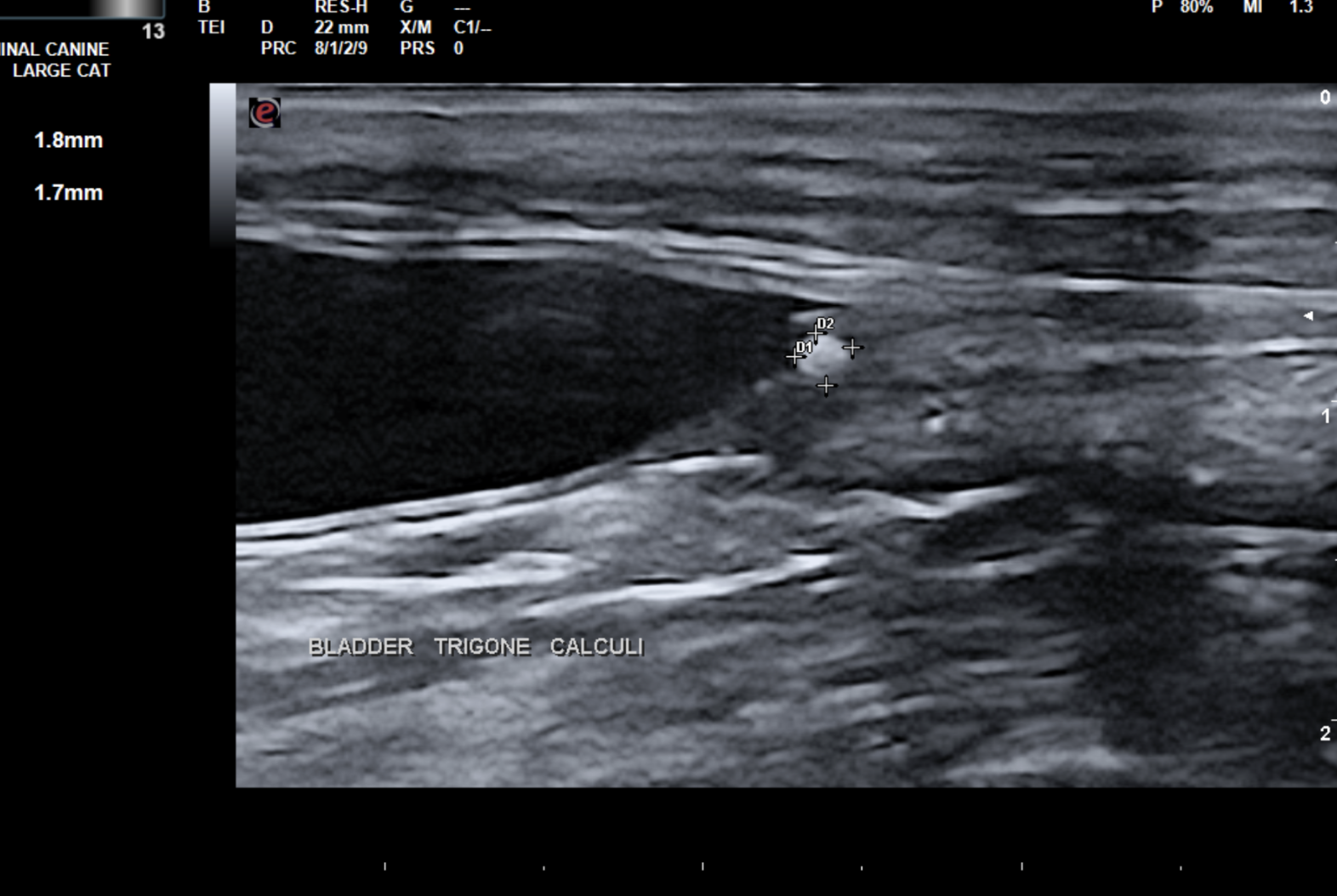
Image 2: Left ureter just distal to kidney

Image 3- Left Ureter approaching the bladder
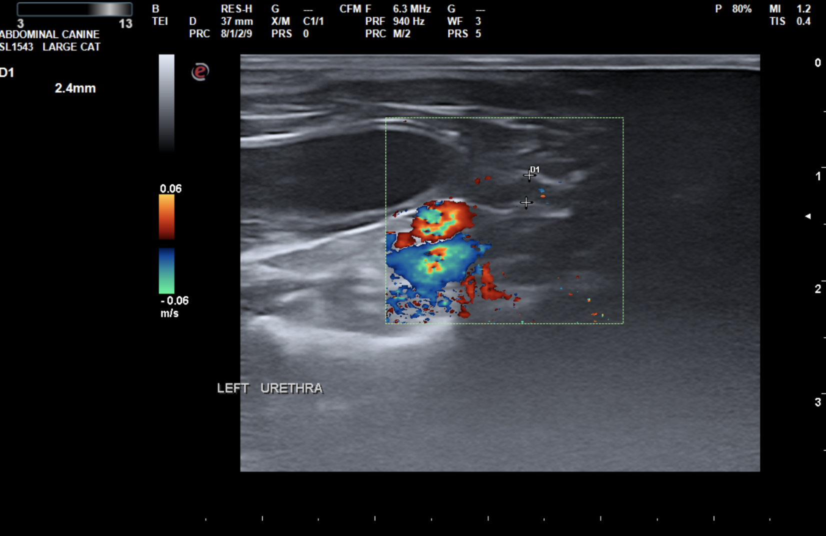
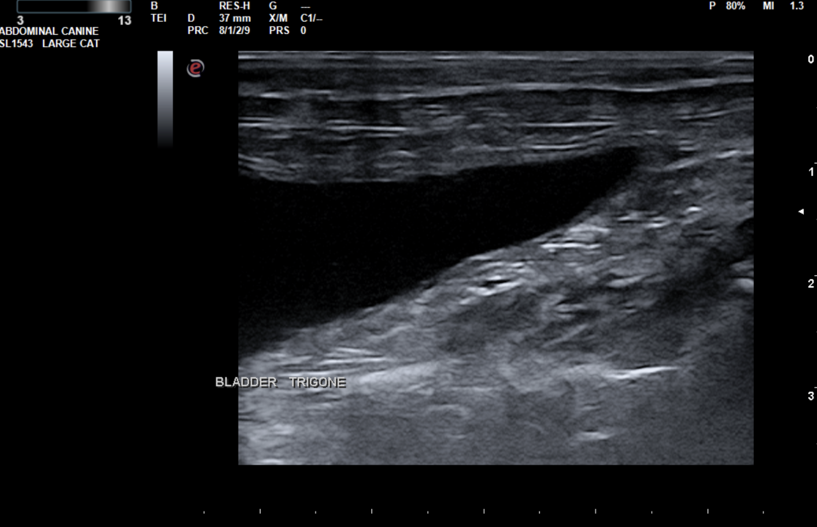
Image 4 - Bladder trigone free of obstruction at 30 and 60 days. No evidence of hydroureter
Diagnosis: Distal ureteral/ureteral papilla partial obstruction with associated mild hydroureter.
CASE OUTCOME:
We are happy to report that this patient spontaneously passed the obstruction and has continued to do well with a healthy appetite. Given the very distal position of the obstruction, lack of significant pyelectasia (indicating likely a partial or resolving obstruction), and the improvement in the patient’s appetite, supportive care and acupuncture were continued. The patient was closely monitored for deterioration as aggressive therapy was not pursued. The patient’s appetite continued to improve. Follow-up ultrasounds at approximately 30 and 60 days showed complete resolution of the obstruction, with no mineralizations or obstructions present in the urinary tract or bladder.
Her CKD will continue to be monitored. There was a mild improvement in renal function after resolution of the obstruction, indicating the likely presence of CKD prior to the development of the obstruction.
PROGNOSIS/DISCUSSION:
The prognosis is good, with some risk of developing future ureteroliths or stricture. It is likely that this patient already had chronic renal disease, which progressed secondary to the ureteral obstruction. Other hematological changes associated with CKD were noted on labs.
Clinical signs of partial or unilateral ureteral obstruction in cats may be as subtle as anorexia or pain. Changes in renal parameters in cats showing signs of pain or anorexia should be investigated with abdominal ultrasound.
In some cases, ureteral obstruction can result in compensatory hypertrophy of the contralateral kidney (not present in this case). All cases of “Big-Kidney, Little Kidney syndrome” should be investigated for signs of ureteral obstruction, independent of the uremic state.
 |  |  |
References:
Kochin EJ, Gregory CR, Wisner E, et al. Evaluation of a method of ureteroneocystostomy in cats. J Am Vet Med Assoc 1993; 202: 257–260.
Lamb CR. Ultrasonography of the ureters. Vet Clin North Am Small Anim Pract 1998; 28:823-848.
Thank you to our friends at Integrative Veterinary Center of Richmond for including us in the care of this lovely patient.