Incidental Abdominal Mass
Patient Information:
- Age: 8 years
- Gender: Spayed Female
- Breed: Terrier Mix
- Species: Canine
History:
The patient initially presented for a new patient exam and annual vaccines. The patient had been doing well at home, aside from gastrointestinal concerns. There is a history of mast cell tumors, and a new lump was recently noted on the right hip. Fine needle aspirates of the dermal mass indicated a mast cell tumor. A lumpectomy was scheduled, with pre-op labs showing normal results. However, on the day of surgery, an abdominal mass was palpated during the pre-op physical exam. Radiographs confirmed a mass in the mid-ventral abdomen. Ultrasound and fine needle biopsy (FNB) of the mass were recommended to rule out metastasis of the mast cell tumor.
Ultrasound Findings:
Mid-Abdominal Left-Sided Mass: A rounded, hypoechoic mass (~2.5 x 1.7 cm) closely associated with and attached to a loop of jejunum and the body wall. The mass is minimally vascular and does not create a distal acoustic shadow. It mildly compresses the attached loop of the small intestine without causing a full obstruction.
Pancreas: Enlarged with scalloped margins and coarse, hyperechoic echogenicity. No focal lesions are observed. The peripancreatic fat has normal echogenicity.
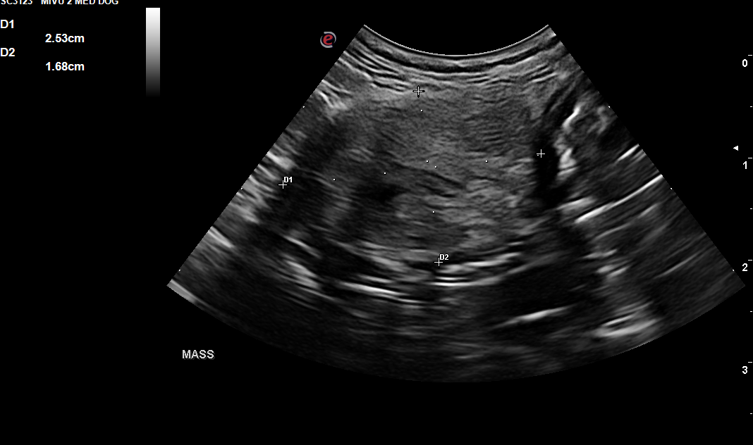
Image 1: Left abdominal mass with adhesions to surrounding jejunum and body wall.
Cytologic Diagnosis and Findings:
Ultrasound-guided fine needle biopsies of the abdominal mass were collected and submitted for cytology. The mass was firm and difficult to penetrate with a needle.
Microscopic Description:
The sample is of low cellularity. Most slides contain low numbers of erythrocytes. One slide contains low numbers of variable intact mononuclear cells, including mixed lymphocytes, neutrophils, and macrophages. Occasional plasma cells are present throughout. Rhomboid amber-colored hematoidin crystals are observed on scanning. Macrophages often contain phagocytosed dark green to black pigment consistent with hemosiderin. No infectious agents are identified.
Diagnosis:
Previous hemorrhage and mixed inflammation
Cytology Comments:
Cytologic findings are consistent with sampling from an area of previous hemorrhage and mixed inflammation. The overall low cellularity of the sample suggests this is likely not fully representative. A biopsy with histopathology is recommended for further characterization.
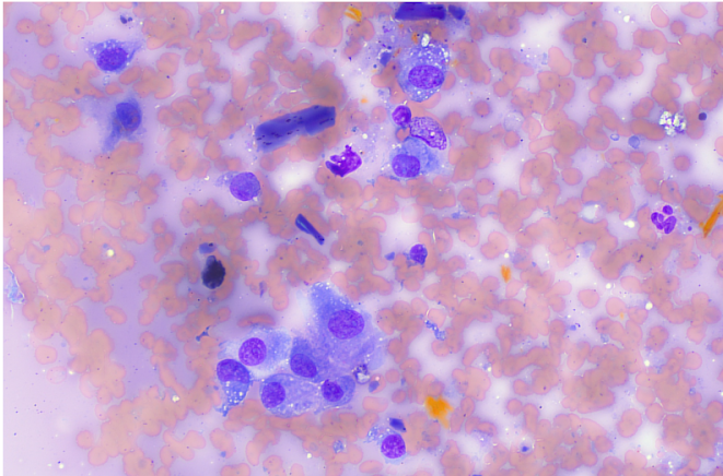
Image 2: Cytology
Further Investigation:
Exploratory surgery was performed to remove the abdominal mass for biopsies. Numerous adhesions to the body wall and intestine were identified during the procedure. Resection and anastomosis of a loop of jejunum were necessary.
Recheck Ultrasound:
A recheck ultrasound was requested 7 days post-exploratory surgery due to reduced appetite, vomiting, and diarrhea.
Findings:
- Jejunal Resection and Anastomosis Site: Suture visible, intact with no evidence of leakage, adhesions, stricture, or tumor growth.
- Pancreas: Findings are moderate and progressive. DDx: chronic pancreatitis, historical pancreatitis, or acute pancreatitis (less likely).
- Mesentery: Findings are moderate. DDx: normal post-laparotomy change, peritonitis (inflammation), paraneoplastic reaction, infection, fibrosis, or other causes.
- Ascites: Finding is trace. DDx: transudate, hemorrhagic, or exudate.
- Lymph Nodes: Findings are mild. DDx: reactive, infection, IBD, infiltrative neoplasia (lymphoma, mast cell, or other), or metastatic neoplasia.
- Spleen: Findings are mild. DDx: reactive hyperplasia, extramedullary hematopoiesis (EMH), infiltrative neoplasia, or benign nodular regeneration.
- Intestines: Findings are mild. DDx: enteritis, inflammatory bowel disease, food allergy/intolerance, or infiltrative neoplasia (unlikely).
All findings indicated normal post-operative changes.
Image 3: Recheck 7 day post operative ultrasound image of resection and anastomosis at mass removal site.
Plan:
Pending results of histopathology, supportive care was continued.
Histopathology Results:
Microscopic Findings: Focal nodular pyogranulomatous to granulomatous steatitis with granulation tissue, fibroplasia, and foreign material.
Comments:
An overt neoplastic process was not observed in the sections examined. The marked inflammatory response and fibrosis are consistent with foreign material resembling plant or synthetic fiber. A differential diagnosis to consider is gossypiboma (retained surgical sponge). The large amounts of dense collagen fibers at the periphery are consistent with chronicity.
In a retrospective study, 12 out of 13 cases had an excellent outcome following the removal of a retained surgical swab (sponge). Preoperative diagnosis was difficult, with a definitive diagnosis made in only one case via diagnostic imaging involving a swab containing a radio-opaque marker. Complications reported in older literature included osteomyelitis and malignant transformation with chronic retention.
Discussion:
Discussion with the owners revealed that the only previous surgery the patient had undergone was an ovariohysterectomy (OVH) performed in another state five years earlier. The incidental finding of a gossypiboma is uncommon and may be underreported. Most dogs with retained surgical gauze present with symptoms such as a palpable mass, vomiting, abdominal pain, weight loss, diarrhea, lethargy, fever, abdominal distension, anorexia, and, in some cases, dermal fistula development. It is unclear if these clinical signs were noted in this patient immediately following the OVH five years ago.
Ultrasonographically, gossypibomas in dogs are seen as well-defined hypoechoic masses with an irregular, hyperechoic center often containing strongly hyperechoic and shadowing foci. The mass in this patient exhibited no acoustic shadowing, possibly due to its chronicity.
Outcome:
After exploratory surgery and the recheck ultrasound, post-operative supportive care was continued. The patient showed steady improvement and made a full recovery.
References:
Louvet A, Duconseille AC. Imaging Diagnosis - Ultrasound Uncommon Features of an Abdominal Gossypiboma in a Dog. Vet Radiol Ultrasound. 2017 Nov;58(6):E68-E70. doi: 10.1111/vru.12453. Epub 2016 Nov 20. PMID: 27866380.
Merlo M, Lamb CR. Radiographic and Ultrasonographic Features of Retained Surgical Sponge in Eight Dogs. Vet Radiol Ultrasound. 2000 May-Jun;41(3):279-83. doi: 10.1111/j.1740-8261.2000.tb01491.x. PMID: 10850880.
Diagnostic Imaging Features of Abdominal Foreign Body in Dogs: Retained Surgical Gauze. J Vet Clin 2011;28:94-100.
Sonographer:
Emily Evans, DVM
Thank you to Sycamore Veterinary Hospital and Eastern Vet Path for collaborating with us on this case.